Content:
◉ Introduction
The beginning of microscopy dates back to the beginning of the 17th century, and since then it has undergone enormous evolution both in terms of performance and in its areas of application.
The microscopic examination, also known as direct examination, is an indispensable tool in an analytical laboratory. Once mastered, this technique can quickly provide valuable information for diagnostic guidance.
In this article, we will limit ourselves to the basic uses of the optical microscope in the medical analytical laboratory, including handling, techniques and different applications.
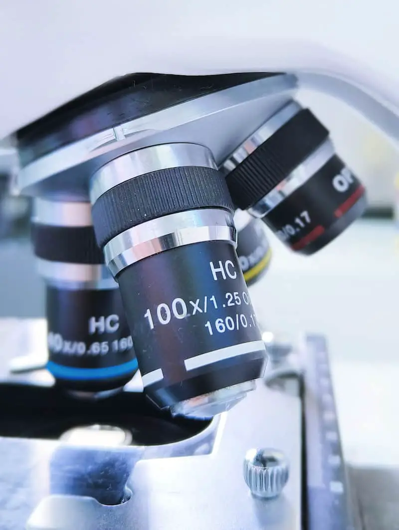
◉ Focusing with an Optical Microscope
Focusing in a microscope involves adjusting the distance between the microscope objective and the sample to get the sharpest image of what you are observing. Here are the general steps for tuning:
- Place your sample on the microscope stage and secure it with the clamps. Start with the low magnification objective (X 10).
- Use the control knobs, usually adjustment screws on the sides of the microscope, to adjust the height of the stage and bring the sample close enough to the objective.
- Look through the eyepiece and use the macrometric adjustment screws to achieve a rough initial focus. Turn these screws clockwise to lower the platen and counterclockwise to raise it.
- Once you have a rough image, use the micrometer adjustment screws to fine-tune the focus. These screws allow for more precise and subtle adjustments.
- If you need to change lenses to achieve higher magnification, rotate the objective turret to select the desired objective. You may need to adjust the focus slightly after changing lenses.
- With regular practice and experience, you will be able to use a high magnification objective directly.
◉ Microscopic Examination Wet Mount
It is a rapid, easy, and cost-effective technique that allows visualization of the sample under a microscope without any preparation (neither fixation nor staining).
The wet mount allows visualization of the various components of the sample, but its greatest value lies in its ability to keep bacteria and parasites alive, allowing optimal observation of their numbers, movements and of their arrangement.
It can be performed directly from the pathological product (urine, cerebrospinal fluid, etc.) or from a bacterial suspension. Various supports can be used, such as slides and coverslips, Malassez cells, and Nageotte cells.
Observation is generally done with a ×40 objective with an almost closed aperture to increase contrast. It is advisable to wait a few moments until the fluid movements stop.
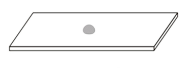
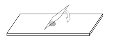
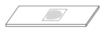
1- Wet Mount Between Slide and Coverslip
Wet mount is a common, easy, quick and widely used practice in bacteriology laboratories. Here are the main steps:
- Place a drop of your sample on a slide. This sample can be a biological liquid (for example urine), a biological suspension (for example a small quantity of stool diluted in sterile water) or a bacterial suspension (a few bacterial colonies dissolved in sterile water).
- Cover the drop with a coverslip.
- Let it rest on a horizontal surface for approximately 10 minutes.
- Observe the sample under a microscope with ×40 magnification.
Homogenize the suspension
Take a drop of the bacterial suspension and place it on a clean slide
Place the coverslip,
Attention! The liquid must not overflow.
Depending on the type of sample and the purpose of the sample, wet mount examination can detect the presence or absence of several biological components such as:
- Bacteria: to assess their shape, arrangement and possible mobility.
- Parasites: to assess their shape, mobility, etc.
- Yeasts.
- Epithelial cells: different types of cells such as renal, bronchial, etc.
- The different variants of red blood cells.
- White blood cells.
- Crystals
- ...etc.
Exemples
Video 01: False bacterial mobility
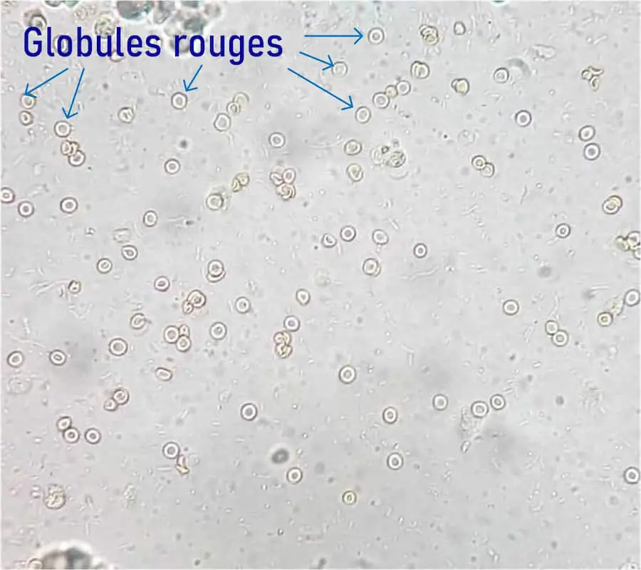
Photo 01: Wet mount of urine, observed under a microscope with a magnification of X40, showing the presence of numerous red blood cells.
2- Hemacytometer Cells
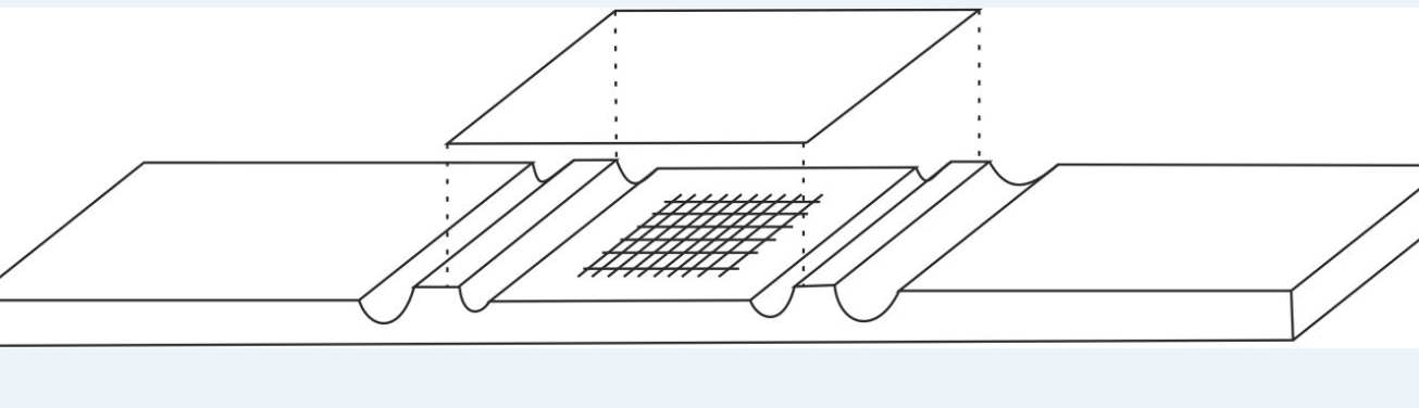
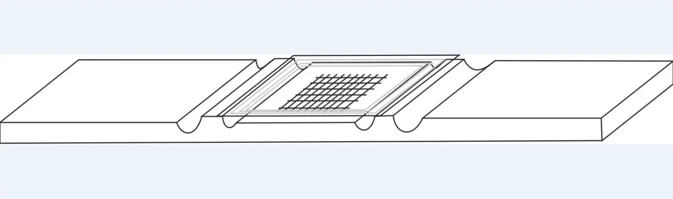
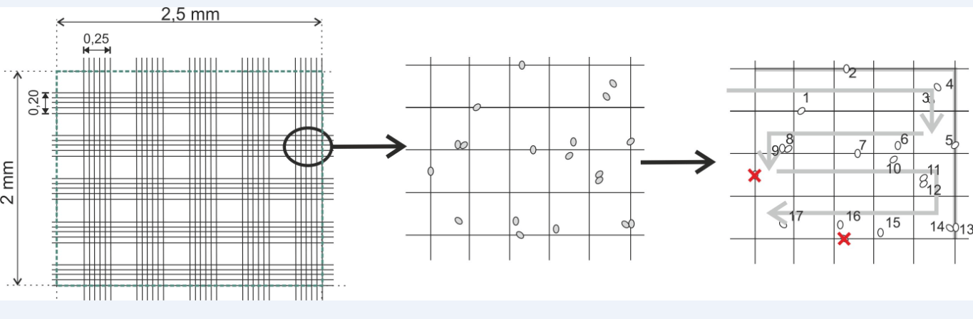
The counting of different elements of biological liquids is possible outside of any automatic machine thanks to the use of calibrated glass cells called hemocytometers.
These are thick glass blades hollowed out with channels, presenting a particular grid on the slightly lowered central platform. Covered with a coverslip placed on the raised side platforms, they allow, with a very precise and known volume, to count the cells of biological liquids.
There are several types of hemocytometers, all with different counting grids, among the most used are the Malassez cell, the Thoma cell, the Nageotte cell and the Neubauer cell.
To obtain a count close to reality, it is important to:
- Do not scratch the grid when cleaning the cell.
- Securely mount the cover onto the cell
- Deposit the sample drop to be counted correctly, as described below
- Allow the particles to be counted to settle before counting.
2. 1 Manipulation
- Place the counting cell on a horizontal surface.
- Stick the cover slip on the cell by moistening both edges with a small, clean, wrung-out damp cloth.
- Slide the coverslip across the width of the cell and check for good adhesion between the cell and the coverslip.
- Homogenize the liquid to be analyzed (or its dilution).
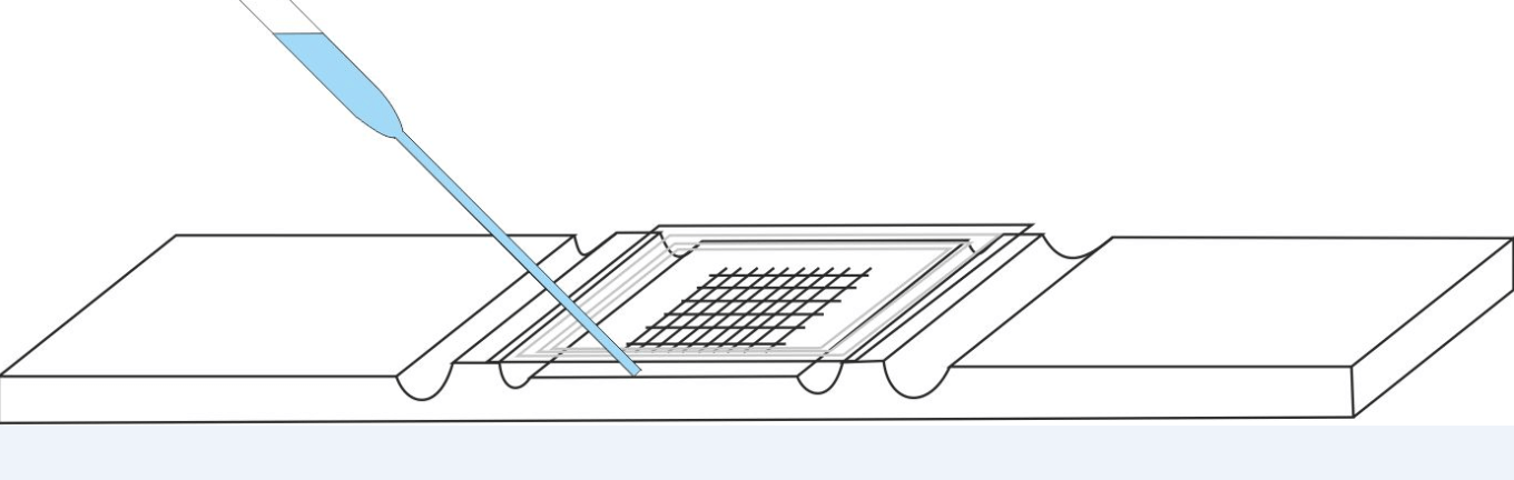
- Between the cell and the coverslip, allow a drop of the sample to be counted to enter, by capillary action, using a Pasteur pipette. The drop must not overflow into the channels of the cell and must completely cover the entire grid surface of the cell at once.
- Wait at least 5 to 10 minutes, keeping the cell horizontal (the cellular elements must sediment).
➊
➋
➌
➍
Note:
- If the fluid to be analyzed is hemorrhagic, it is recommended to lyse the red blood cells before counting the other cells by diluting the hemorrhagic fluid by half using a 0.5% solution of acetic acid in 'water.
- If the liquid to be analyzed has a high number of cells, it should be diluted in physiological saline as necessary, for example 1/2, 1/5, 1/10 or more.
2. 2 Counting
- If counting is carried out on diluted liquid, do not forget to multiply the number of elements counted by the dilution rate.
◉ Microscopic Examination After Staining
The coloring of biological products is a very common technique in medical biology. It allows you to provide more details or even make visible elements that are invisible when fresh.
There are several types of coloring, with different production procedures. The choice depends on the type of sampling and the element sought. Roughly, they can be classified into simple (non-differential) colorations and differential colorations
The common first step is usually taking a smear and fixing it. Then, after coloring, the reading is done at a magnification of X100 with an open diaphragm.
1- Simple staining
Simple stains, or direct staining, are quick-to-perform procedures that involve adding a single dye to the smear, leaving it to act, then washing, drying and observing under a microscope. They make it possible to determine the presence and morphology of biological elements such as bacteria, cells, etc.
Simple coloring uses basic dyes such as methylene blue, safranin, crystal violet, malachite green, etc.
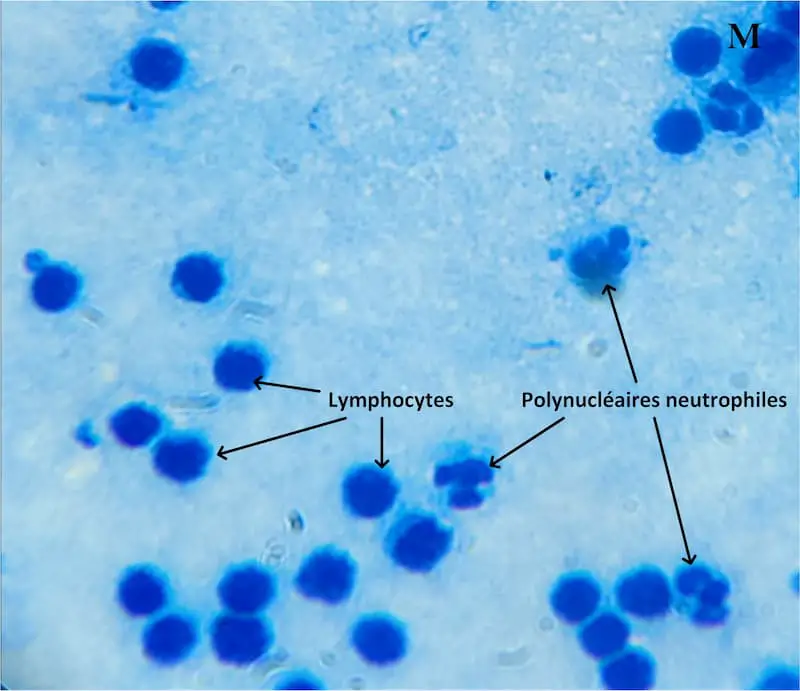
Photo: Methylene blue (basic) stains the nuclei of leukocytes (acidic), which makes it possible to differentiate between lymphocytes and polymorphonuclear cells.
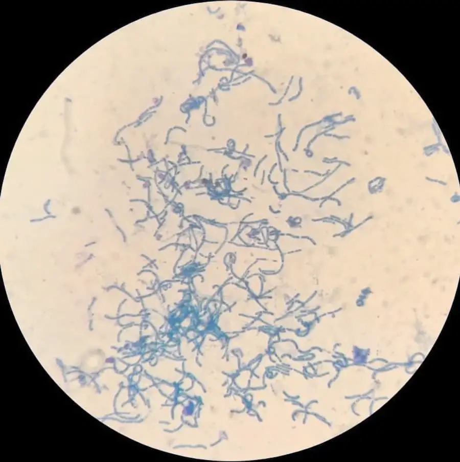
Photo: Lactobacillus stained with methylene blue
2- Differential staining
Differential staining involves using multiple dyes to differentiate between various types of cells, structures, and microorganisms.
There are a number of differential staining methods, such as Gram staining, considered the gold standard in bacteriology, which allows bacteria to be differentiated into Gram positive and Gram negative. Ziehl-Neelsen staining makes it possible to detect and differentiate tuberculosis bacilli from other bacteria.
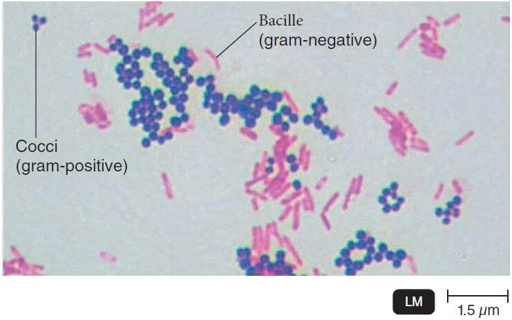
Photo: Gram stain showing Gram-positive cocci and Gram-negative bacilli
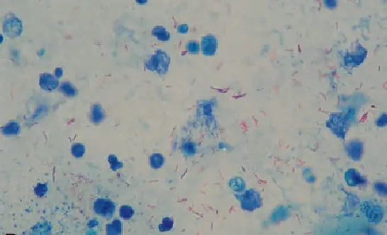
Photo: Koch's bacillus stained by Ziehl-Neelsen stain
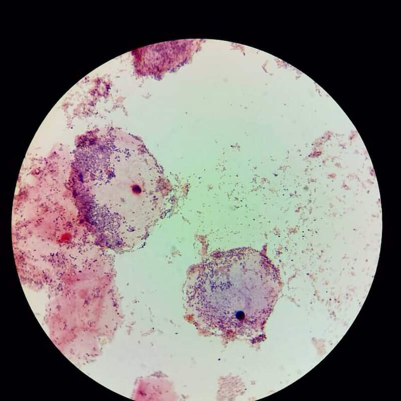
Photo: A Gram-stained V.swab, observed under a microscope with a magnification of X100, showing the presence of Clue-cells
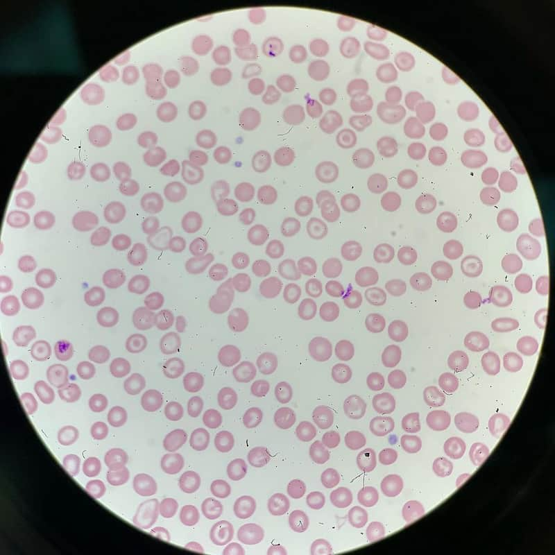
Photo: Blood smear stained with MGG, observed under a microscope with a magnification of X100
◉ Conclusion
In conclusion, microscopic examination proves to be an essential technique in the medical analysis laboratory. It allows detailed observation of biological samples, providing valuable information for diagnosis and patient management.
It is essential to emphasize the importance of rigor and precision in carrying out this review, as well as the need to take into account the various sources of potential errors.