Contents:
Methylene blue staining
Ⅰ.Introduction
In 1886, methylene blue was used for the first time in the medical field. Dr Paul Ehrlich discovered a strange phenomenon in an experiment: Methylene blue stained living neurons blue, and it has the same effect on Plasmodium (a parasite that causes malaria) in human blood.
Currently, it is widely used in chemical indicators, dyes, biological dyes and drugs Methylene blue stain is a fast, economical and commonly used stain.Ⅱ. Methylene blue: Properties
Methylene blue, or methylthioninium chloride, is an organic, odorless crystalline solid soluble in water and, to a lesser extent, in ethanol. In its pure state, it comes in the form of a dark green powder, it is also found commercially as a double salt with zinc chloride, brown in color.
Methylene blue is an oxidation-reduction agent (depends on its redox state: it is colorless in the reduced state, but is blue in the oxidized state).
Ⅲ. Preperation
- Dissolve 0.3 to 0.5 g of methylene blue chloride in 100 ml of distilled water
- Mix the crystals and the liquid until the crystals are completely dissolved.
- Filter a small part of the reacted liquid before each use.
- The ideal is to be able to let the methylene blue oxidize for a few weeks before use in order to be able to obtain a kind of "metachromatic" coloration. To do this, stopper the bottle with a piece of carded cotton and not with the standard airtight stopper.
◈ Methylene Blue solution according to Loeffler : Methylene blue: 0.30 g + Ethyl alcohol 95% : 30.0 ml + Distilled water 100.0 ml
Ⅳ. Principal
Since cells usually have negatively charged cell walls, the chromophores of basic, positively charged dyes tend to stick to cell walls, making them positive spots.
Methylene blue stain is a simple stain where a single dye is used to emphasize particular structures in the sample, shape (the size and arrangement of bacteria). The organisms in a sample will be the same color, even if the sample contains more than one type of organism.
◈ In some cases, the methylene blue stain gives better results than the Gram stain. Among these cases :
- Methylene blue (according to Löffler) has traditionally been used to stain Corynebacterium diphtheriae for the observation of metachromatic granules.
- Y. pestis , When stained with methylene blue, it presents an intense staining at each end of the bacillus, called bipolar staining, which gives it a "safety pin" appearance.
- Burkholderia pseudomallei : typical bipolar "safety pin" appearance.
- In cerebrospinal fluid, allows better contrast between Gram-negative bacteria, Neisseria meningitidis and Haemophilus influenzae and the background
- Haemophilus ducreyi stained with methylene blue, allowing to observe the typical "bicycle chain" appearance.
- Respects cell structure better than Gram.
- It is also used as a counterstain in several other stains (method of Moeller, Macchiavello, Stamp, Ziehl-Neelsen ..)
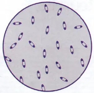
y.pestis "security pin" aspect
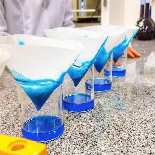
Ⅴ. How to stain with methylene blue
- After performing the bacterial smear (smearing, fixing)
- Place the slide on a staining rack and flood it with methylene blue. Leave on for 1-3 minutes
- Gently wash the slide with distilled water, drain off excess water, blot (do not rub) with absorbent paper and allow slides to air dry completely.
- Examine with the x100 immersion objective (with a drop of oil) with strong illumination (diaphragm open)

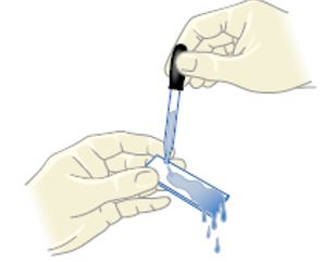
Pour a methylene blue solution onto a smear
After 1-3min rinse with tap water

Dry between two sheets of blotting paper then observed by immersion
Checking for leukocytes in diarrheal stools
Place a small drop of faecal mucus or liquid stool on a microscopic slide. Add an equal volume of methylene blue (Loeffler blue). Mix with a small wooden stick; cover with a strip. Leave the coloring to take place for 2 to 3 minutes. Examine with the dry objective (× 40 or × 60).
Ⅵ. Interpretation
The stainable structures appear blue, the staining makes it possible to detect the morphology of these structures, their grouping mode and in some cases the cell type
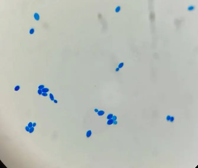
Yeasts stained with Methylene blue
References:
- Sigma-Aldrich- 50484 Methylene Blue Solution
- protocols.io- Methylene Blue staining
- Book : Textbook of diagnostic microbiology 4th ED
- Microbiology - OpenStax
- Book : Bactériologie médicale Techniques usuellese
- Book : Clinical Microbiology Procedures Handbook 4ed
- Book : Laboratory Manual and Workbook in Microbiology Applications to Patient Care 7th Edition
- Dr. vikassaini : Yersinia, Pasteurella and Francisella
- microbe-canvas : Methylene Blue staining
- Himedia : Methylene Blue (Loeffler's)
- Microbiología Ambiental : COLORACIÓN DE AZUL DE METILENO DE LOEFFLER (coloración de gránulos metacromáticos):