Content :
We examine every vaginal sample we receive for the presence of clue cells due to their significant importance in identifying bacterial vaginosis. This article focuses on the characteristics of clue cells, their detection and explores their clinical significance.
◉ What are Clue Cells ?
Clue cells are vaginal epithelial cells that have the characteristic appearance of having a grainy border (speckled appearance) when viewed under a microscope.
First described by Gardner and Dukes, the presence of clue cells, along with other clinical criteria, can help diagnose bacterial vaginosis.
The elevated pH that occurs in bacterial vaginosis facilitates the adhesion of certain bacteria 'Gardnerella vaginalis', which then produce cytotoxins and a biofilm which exfoliates, resulting in a distinct appearance.

Clue cells under microscope
◉ Diagnosis of clue cells
To detect clue cells, a healthcare provider will perform a vaginal exam and collect a sample of vaginal discharge using a sterile swab for microscopy observation.
- Place the sample on a clean microscope slide by rolling the swab back and forth.
- Let it dry, then perform a Gram stain.
- Examine the slide under a microscope using oil immersion and a high magnification objective lens (e.g. 40x or 100x).
- Look for vaginal cells that have small, coccobacilli-shaped bacteria adhered to their surface.
◈ Note:
- Microscopy observation should be used in conjunction with other diagnostic methods and clinical evaluation
- To be meaningful for bacterial vaginosis, more than 20% of epithelial cells on the Pap smear must be clue cells (60-80% sensitivity and 99% specificity ).
◉ What does it mean if you have clue cells ?
Normally, the vagina contains a balance of different types of bacteria that help maintain a healthy environment. The presence of clue cells in a sample of vaginal secretions indicates an abnormal overgrowth of certain types of bacteria and depletion of protective bacteria such as lactobacillus, this is a hallmark of bacterial vaginosis, a common vaginal infection.
This bacterial infection is characterized by:
- Foul fishy odor
- Abnormal vaginal discharge
- An increased vaginal pH > 4.5
◉ What are clue cells caused by?
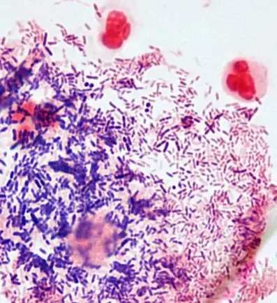
Gardnerella vaginalis is a type of bacteria that is commonly associated with bacterial vaginosis. It is a Gram-variable bacilli or coccobacilli ; catalase and oxidase negative; sodium hippurate hydrolysis positive; acid production from glucose, maltose, and sucrose (variable)

Gardnerella vaginalis : Gram-variable rods
◉ Is it normal to have some clue cells ?
It may indicate the presence of BV. However, the interpretation should be made in conjunction with other clinical and laboratory findings, as the presence of clue cells alone is not sufficient to diagnose BV.