Sommaire :
◉ Introduction
Most bacteria are colorless, so they generate little contrast in the microscope field. Therefore, to see bacteria under the microscope, it is necessary to apply color using a staining reagent. Once stained, bacteria can be observed and studied in terms of their shape, size and arrangement.
A bacterial smear is a thin layer of bacteria placed on a glass blade and «fixed», by heat or other, partly to ensure that they remain attached to the glass, for coloring. It can be prepared from a solid medium or broth
Then a coloring is applied. The surface and cytoplasm of bacterial cells , DNA, RNA, proteins and chromatin are negatively charged (acid) to color them a positively charged (basic) dye is applied (methylene blue, basic fuchsin, violet crystal, saffron, malachite green).
◉ General considerations
There are some important things to consider when preparing a smear for staining:
- The bacteria should be evenly and lightly dispersed.
- Bacteria should be firmly attached to the slide so that they are not washed out during staining procedures.
- It is very easy to not know which side of the slide the smear is on. Be sure to label the far edge of the slide.
- Allow your slide to air dry before you heat set it (bacteria will boil and cell morphology will be lost)
◉ Performing smears
❶ Smear spread
Degrease the surface of a glass slide with alcohol on a Bunsen burner, place it on a staining support with the degreased surface upwards, leave to cool::
From cultures
- If you are working from a solid medium, place a drop of water in an area on the slide. If you are using medium broth, no additional liquid is needed.
- With your inoculation loop, completely mix the sample (bacterial colonies) with the water and spread the mixture to cover approximately half of the total surface of the slide.
From swabs (sample)
- Smears should not be prepared from a swab after it has been used to inoculate culture media. Ideally, if the sample can only be taken from swabs, two swabs are submitted.
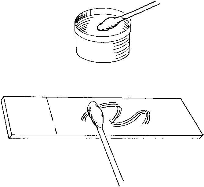
- Swab smears are prepared by rolling the swab back and forth (The swab should never be rubbed, the smear elements could be broken) over adjoining areas of the glass slide to deposit a thin layer of sample material. This preserves the morphology and relationships of microorganisms and cellular elements.

From thick liquids or semi-solids
- Swabs can also be used as a tool for preparing smears from thick or semi-solid liquid samples such as feces.
- The swab is immersed in the sample for several seconds, then used to prepare a thin layer of material on the glass slide for staining and visualization. This swab method of preparation is adequate but may produce less desirable results than other methods.

❷ Drying and Fixing of Smears
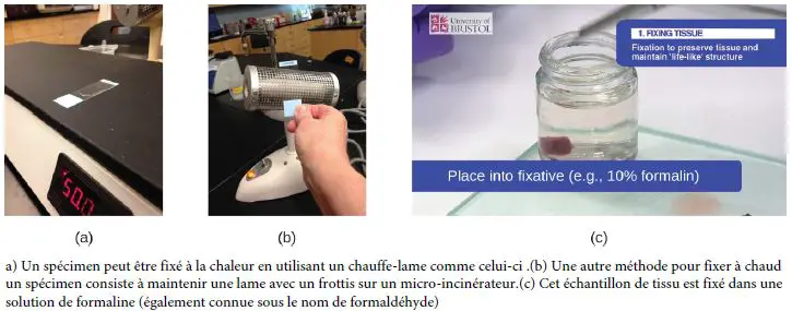
- Drying must precede fixing. Should be done as much as possible at laboratory temperature, if necessary by placing the slide on a heating plate set at 37 °, or by keeping it in hot air above the pilot burner of a Bunsen burner.
Note: Precautions must be taken to avoid extreme heat because deformation of the cell and splashing may occur (alteration of certain constituents, in particular the bacterial wall).
- “Fixation” of a sample refers to the process of attaching cells to a slide. Fixation is often obtained either by heating (eg by quickly passing the blade three times through the Bunsen burner), or by chemical treatment of the sample. In addition to attaching the sample to the slide, the attachment also kills microorganisms in the sample, arresting their movement and metabolism while preserving the integrity of their cellular components for observation.
