✑ Dr. Belouahad loqman
Summary :
◉ Overview
The ability of bacteria to ferment lactose depends on two enzymes, permease and beta-galactosidase .
Permease allows lactose to enter the bacterial cell wall, where it is then broken down into glucose and galactose by beta-galactosidase . Glucose and galactose can then be metabolized by bacteria.
However, some organisms lack permease and appear to be late or non-fermenters.
☰ The ONPG test is used to detect the enzyme β-galactosidase , present in late lactose fermenters (late lactose fermenters are very difficult to distinguish from non-lactose fermenters because both appear under form colorless colonies on MacConkey agar ).
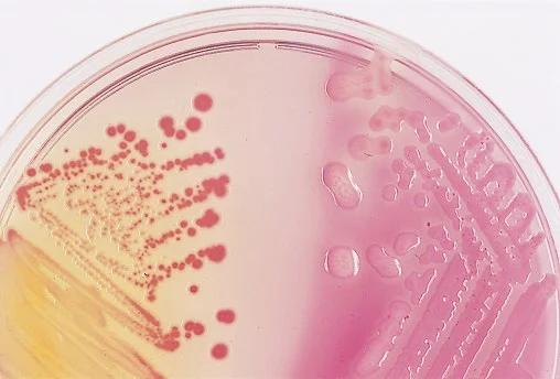
Enterobacter spp. (colonies in the left) are often late lactose fermenters, and so colonies may appear colorless to light pink.
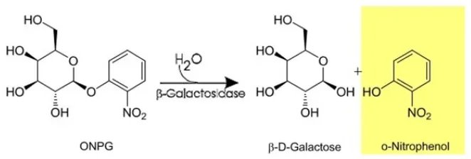
ONPG (O-Nitrophenyl-β-D-galactopyranoside) is structurally similar to lactose (i.e., ONPG is a lactose analogue) except that orthonitrophenyl has been substituted for glucose.
◉ Principle of ONPG test
O-nitrophenyl-beta-D-galactopyranoside (ONPG) is an artificial substrate structurally similar to lactose except that glucose is substituted with an o-nitrophenyl group. Unlike lactose, ONPG is able to enter the bacterial cell without the presence of permease.
During hydrolysis, by the action of the β-galactosidase enzyme, ONPG splits into two residues, galactose and o-nitrophenol. ONPG is a colorless compound: O-nitrophenol is yellow, providing visual evidence of hydrolysis.
◉ Procedure of ONPG test
Bacteria grown in media containing lactose (to induce production of the enzyme galactosidase), such as Kligler Iron Agar (KIA) or Triple Iron Sugar Agar (TSI), produce optimal results in the ONPG assay.
☰ In the broth test method, the organism is taken from a medium containing a high concentration of lactose and is inoculated into the ONPG broth. If the body has beta-galactosidase, the enzyme will split the beta-galactoside bond, releasing o-nitrophenol which is a yellow colored compound. This indicates a positive test.
☰ In the disc method, the test organism is taken from a medium containing a high concentration of lactose. A dense suspension (turbidity equivalent to a 3 McFarland) is prepared. An ONPG disc is added to 0.5 ml of the suspension. If the body has beta-galactosidase, the enzyme will split the beta-galactoside bond, creating a yellow color change in the suspension.
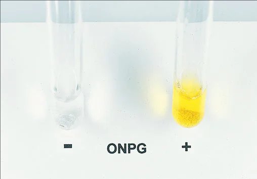
☰ Organisms with strong beta-galactosidase activity may produce a positive reaction a few minutes after inoculation of the ONPG medium; other organisms can take up to 24 hours.
◉ ONPG positive bacteria | ONPG negative bacteria
| O.N.P.G. POSITIVE (yellow color) | O.N.P.G. NEGATIVE (colorless) |
| E. coli, Shigella dysenteriae 1, Certain strains of S. dysenteriae of other serotypes and of S. boydii, Salmonella sub genus III (S. arizonae), Salmonella sub genus II *, Citrobacter, Levinea malonatica, Levinea amalonatica **, Klebsiella (except K. rhinoscleromatis), Enterobacter, Hafnia **, Serratia marcescens **, Serratia liquefaciens **, Serratia plymuthica, Serratia rubidaea, Erwinia carotovora, Yersinia **, Vibrio cholerae **, Plesiomonas | Alcalescens dispar, Other Shigella, Salmonellasous genus I and IV, Edwardsiella, Proteus, Providencia, Vibrio parahaemolyticus |
* Late, after 24 hours of incubation. ** Hydrolyze O.N.P.G. thanks to an enzyme other than β-galactosidase stricto sensu, i.e. inducible by Isopropyl-beta-D-thiogalactopyranoside (I.T.P.G.)
NB: The test at the O.N.P.G. may have an interest in other bacterial genera; for example, in Pseudomonas aeruginosa, strains of serogroup O: 11 are with rare exceptions, O.N.P.G. (+) unlike those of other O groups, in Neisseriaceae, N. lactamica is the only O.N.P.G. (+).