Content :
◉ Overview
To survive, organisms must rely on defense mechanisms that allow them to repair or escape oxidative damage from hydrogen peroxide (H2O2). Some bacteria produce the enzyme "Catalase" which facilitates cellular detoxification.
The Catalase neutralizes the bactericidal effects of hydrogen peroxide and its concentration in bacteria has been correlated with pathogenicity
◉ Why catalase test is done ?
Detection of the presence of catalase in bacteria is essential to differentiate catalase-positive Staphylococcaceae and Micrococcaceae from catalase-negative Streptococcaceae .
Although it is primarily useful in differentiating between these genera, it is also useful in distinguishing between species belonging to the same genus such as Aerococcus urinae (catalase positive) and Aerococcus viridians (catalase negative) and gram negative organisms such as Campylobacter fetus, Campylobacter jejuni and Campylobacter coli (catalase positive) from other Campylobacter species (catalase negative)
The catalase test is also useful in differentiating between obligate aerobic and anaerobic bacteria, as the latter usually lack the enzyme. In this context, the catalase test also serves to differentiate between aerotolerant strains of Clostridium, which are catalase negative, of Bacillus, which catalase positive

◉ Principle of the catalase test
◉ The enzyme Catalase is used to neutralize the bactericidal effects of hydrogen peroxide . The Catalase accelerates the decomposition of hydrogen peroxide (H2O2) into water and oxygen (2H2O + O2).
◉ This reaction is evident by the rapid formation of bubbles
◉ Reagents
Use commercially available 3% hydrogen peroxide to test for an aerobic strain of bacteria. Store hydrogen peroxide in the refrigerator in a dark bottle.
For the identification of anaerobic bacteria, a 15% H2O2 solution is required.
◉ Protocol of the catalase test
There are many method variations of the catalase test. These include the slide or drop catalase test, the tube method, semi-quantitative catalase for the identification of Mycobacterium tuberculosis, the thermostable catalase used for the differentiation of Mycobacterium species and the capillary tube method and cover slip.
The most popular method in clinical bacteriology is the slide or drop catalase method, as it requires a small amount of culture and relies on a relatively uncomplicated technique.
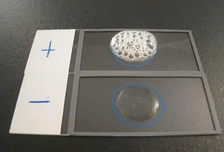
- Place a microscope slide inside a Petri dish.
- Keep the lid of the Petri dish on hand. The use of a Petri dish is optional as the method can be performed correctly without it. It aims to limit aerosols which have been shown to carry viable bacterial cells.
- Using a sterile inoculation loop or wooden application stick, remove a well-isolated colony from a pure culture (18-24 hours incubation) and place it on the slide. microscope.
- Be careful not to take agar. This is particularly important if the cuture was made on blood agar. Red blood cells can cause a false positive reaction.
- Note : If a platinum inoculation loop is used: The platinum wire in the loop may produce a false positive result. This is not the case with Fil Nichrome.
- Using a dropper or Pasteur pipette, place 1 drop of 3% H2O + O2 on the colony. Do not mix.
- Immediately cover the Petri dish with a lid and observe the immediate formation of bubbles (O 2 + water = bubbles).
Note :
- Observing the formation of bubbles on a dark background improves readability.
- Use a magnifying glass to observe weak positive reactions. A microscope can also be used. In this case, place a coverslip on the slide and view under 40x magnification.
- The absence of the formation of bubbles (no catalase enzyme to hydrolyze hydrogen peroxide) represents a negative reaction
2 - Catalase in tube
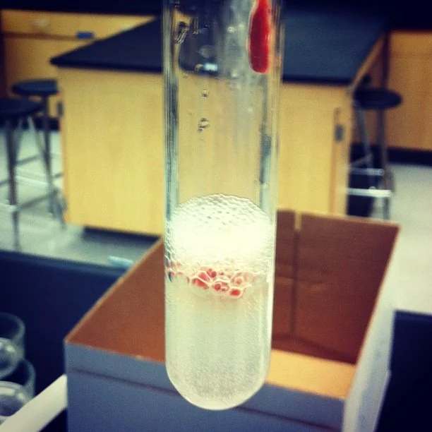
❏ Add 4 to 5 drops of 3% H2O2 to a 12 x 75 mm test tube. Using a wooden stick, take a well isolated colony from the strain to be tested and place it in the test tube.

3 - Tube Method (Slant)
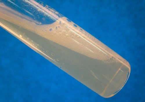
❏ Add 1.0 ml of 3% H2O2 directly to a pure culture heavily inoculated 18-24 hours and grown on a slant of nutrient agar. Place the tube on a dark background and observe the immediate formation of bubbles.

◉ Special cases of the catalase test
1- Catalase test Mycobacterium tuberculosis :
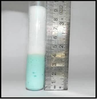
The semi-quantitative catalase test divides mycobacteria into 2 groups, those producing less than 45 mm of bubbles (Positive) and those producing more than 45 mm of bubbles (Negative):
⤠ Usually, Mycobacterium kansasii, Mycobacterium simiae, most scotochromogens, non-photochromogenic saprophytes and fast growing cultures produce more than 45mm of bubbles.
⤠ Mycobacterium tuberculosis, Mycobacterium marinum, Mycobacterium avium complex, Mycobacterium xenopi and Mycobacterium gastri are among those which produce less than 45 mm of bubbles.
Protocol :
1. Inoculate Lowenstein-Jensen with 0.1 ml of a 7-day liquid culture of the test isolate or of an active culture on solid medium collected using a filled loop
2. Include positive and negative control organisms.
3. Incubate the tubes in 8-10% CO2 at 33-37 ° C with the caps loose for 2 weeks.
4. After incubation, add 0.5 ml of catalase reagent (1: 1 mixture of 10% Polysorbate® 80 (REF R21275) and 30% hydrogen peroxide) to the culture.
5. Place the tubes vertically in a rack placed on absorbent paper moistened with disinfectant. The column of bubbles produced may overflow from the tube if the cap is not replaced and tightened quickly enough.
6. Let the tubes stand at room temperature for 5 minutes before measuring them. Measure in millimeters the height of the column of bubbles above the average surface.
Positive - A column of bubbles greater than 45 mm
Negative - A column of bubbles less than 45 mm


2- Catalase test Neisseria gonorrhoeae :
The Superoxol test is a rapid test that can be used for the presumptive identification of Neisseria gonorrhea. It is similar to the Catalase test in which the 3% solution of hydrogen peroxide (H2O2) is replaced with a 30% solution of hydrogen peroxide. Strains of N. gonorrhoeae produce immediate and vigorous bubbles
N. meningitidis and N. lactamica, as well as other species that grow on selective media, generally produce weak and delayed bubbles, although some isolates of N. meningitidis may also produce immediate and vigorous bubbles similar to gonococcus.