Content:
◉ Introduction
The red blood cells, or erythrocytes, are blood cells essential to the proper functioning of the circulatory system. Their main role is to transport oxygen from the lungs to the body's tissues and to transport carbon dioxide from the tissues back to the lungs for elimination.
The ability of red blood cells to perform this function depends not only on their number and hemoglobin content, but also on their shape. Indeed, erythrocytes can present various morphologies, ranging from the normal shape, generally described as a biconcave disc, to abnormal shapes which can result from different etiological processes.
Depending on the disease in question, red blood cells can take on very diverse shapes: spherical, sickle-shaped, crenate, and many others. When the level of these abnormally shaped cells exceeds 10%, we speak of poikilocytosis.
Some changes are typical of certain diseases, such as sickle cell disease. However, proper interpretation of red blood cell morphology is made in conjunction with other observations on the blood smear, complete blood count (CBC), results of other tests and other clinical information.
◉ Appreciation of the shape of red blood cells
Evaluation of the shape of red blood cells is mainly carried out through the examination of the blood smear, a fundamental technique in hematology. The process involves spreading a drop of fresh blood on a glass slide, staining it, and then observing it under a microscope.
Cytological analysis of erythrocytes requires properly spread smears, made from fresh blood, and examined by an experienced biologist.
◉ Normal Red Blood Cell Form
Circulating erythrocytes are the end product of a complex maturation process, which takes place mainly in the bone marrow and is called erythropoiesis.
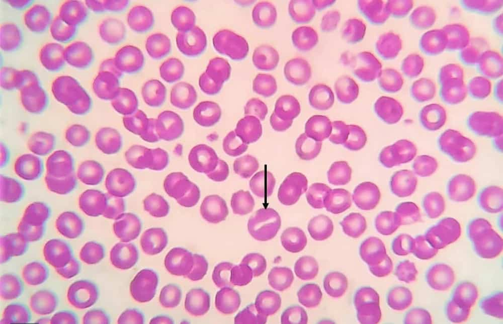
Normal mature red blood cells have a diameter between 6.7 μm and 7.8 μm (6-8.5 µm). They circulate in the blood as biconcave discs, with a central pallor occupying approximately one third of the cell's diameter.
In a healthy situation, the size and shape of red blood cells vary relatively little. The main characteristics that can help identify and differentiate a normal red blood cell from an abnormal red blood cell are:
- Uniform shape, generally biconcave or rounded disc, sometimes slightly oval.
- A central area of paleness that occupies approximately one third of the cell diameter.
- A smooth outline.
- Anucleate, that is to say it does not contain a visible nucleus.
- Absence of visible inclusions inside the cell.
- Absence of variable coloring (polychromasia) in the cytoplasm.
- About the same size as the nucleus of a small lymphocyte.
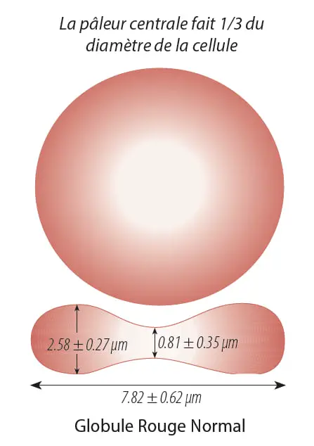
Photo 01: Normal shape of the red blood cell
◉ Poikilocytosis: Abnormal Form of Red Blood Cell
Poikilocytosis refers to changes in the shape of red blood cells. These changes can have several origins:
- Abnormal erythropoiesis, i.e. abnormal production of red blood cells in the bone marrow.
- An intrinsic erythrocyte abnormality, such as a hemoglobin variant, membrane defect, or enzymatic defect.
- Extrinsic causes, for example when erythrocytes are damaged by drugs, chemicals or toxins, by heat or by abnormal mechanical forces.
◉ 1. Spherocytes
Spherocytes are compact, rounded red blood cells, with a regular outline and no pale central area. They have a spherical or almost spherical shape, unlike normal red blood cells which are biconcave disc shaped. Additionally, their diameter is generally smaller than that of normal red blood cells.
Spherocytes can result from genetic defects in the red blood cell membrane, as in hereditary spherocytosis, as well as in transfusion and hemolytic reactions, and also following the action of bacterial toxins, among others.
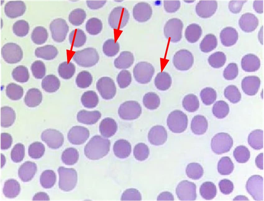
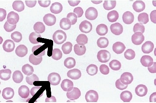
◉ 2. Codocytes (Target Cell)
Codocytes (or target cells) are distinguished by a distinctive ring-like structure: an area of denser staining in the center, surrounded by a pale band, then a circle with normal staining.
This form occurs due to an increase in red blood cell membrane relative to hemoglobin content, resulting in an increased surface-to-volume ratio.
The target cells can be seen in various conditions such as thalassemia, liver diseases, severe iron deficiency anemias and other conditions.
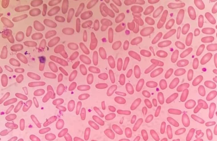
◉ Elliptocyte
The term "elliptocyte" is used to describe elliptical, elongated red blood cells with rounded ends. These cells have a smaller diameter than ovalocytes and have two parallel sides.
Elliptocytes are often present in large numbers in hereditary elliptocytosis.
◉ Schistocytes
Schistocytes, or fragments of erythrocytes, are generally smaller than normal red blood cells and vary in shape. Sometimes they have sharp corners or thorns, or take on an irregular shape.
They are found in many blood diseases, notably megaloblastic and dyserythropoietic anemias, as well as in microangiopathies.
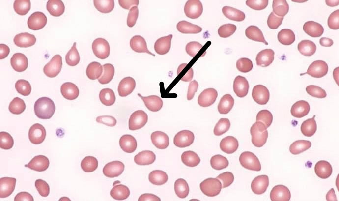
◉ Dacryocytes (tear-drop)
These are erythrocytes shaped like drops, tears or pears, with a pointed end.
This shape can form after erythrocytes containing cellular inclusions pass through the spleen, where splenic macrophages attempt to remove this rigid inclusion, the cell stretches into an abnormal shape.
Although they are strongly associated with myelofibrosis, they can also occur in other disorders such as thalassemia major, bone marrow infiltration, extramedullary hematopoiesis, hereditary elliptocytosis, hereditary pyropokilocytosis, deficiency in severe iron, myelodysplasia, megaloblastic anemia, among others.
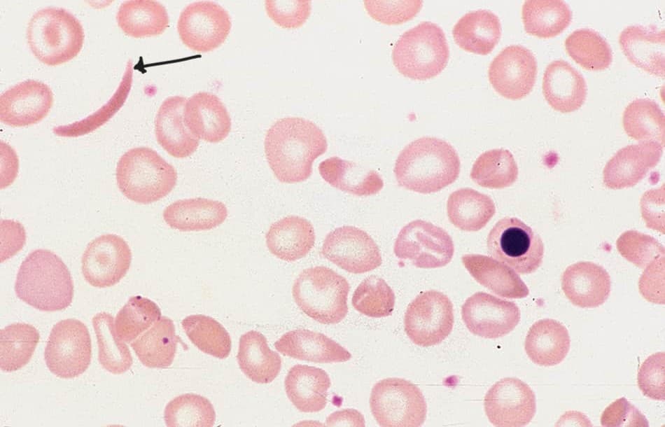
◉ Sickle Cells
Red blood cells have an elongated sickle, crescent, or boat shape, with denser color and pointed ends. This form is characteristic of sickle cell disease (SS, SC, SD , S-Beta Thalassemia).
Sickle cells are usually absent in newborns and are rare in adult patients with a high hemoglobin F percentage.
◉ Keratocytes
Keratocytes have a pair of spicules, usually one or two pairs, separated by a segment of concave semicircular membrane, giving them the appearance of horns. The term "horn cell" is sometimes used to describe keratocytes.
They arise from the ablation of Heinz bodies by macrophages, or from traumatic cellular damage and blister cell rupture.
They are mainly present in G6PD deficiency anemia and angiopathic hemolytic anemia.
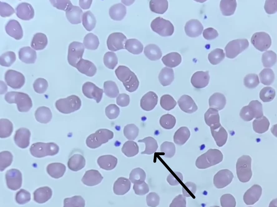
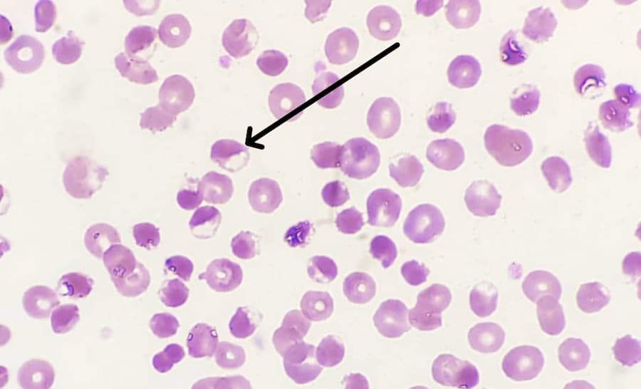
◉ Stomatocyte
Stomatocytes are erythrocytes which have a mouth-shaped slit in their clear central region, often compared to a "fish mouth".
The presence of stomatocytes is seen in various conditions such as hereditary stomatocytosis, Southeast Asian ovalocytosis, excessive alcohol consumption, liver and biliary abnormalities, obstructive pulmonary diseases, as well as in certain cases of myelodysplastic syndromes. They can also be observed as a secondary artifact of technical manipulations.
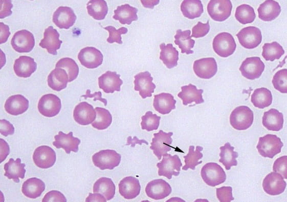
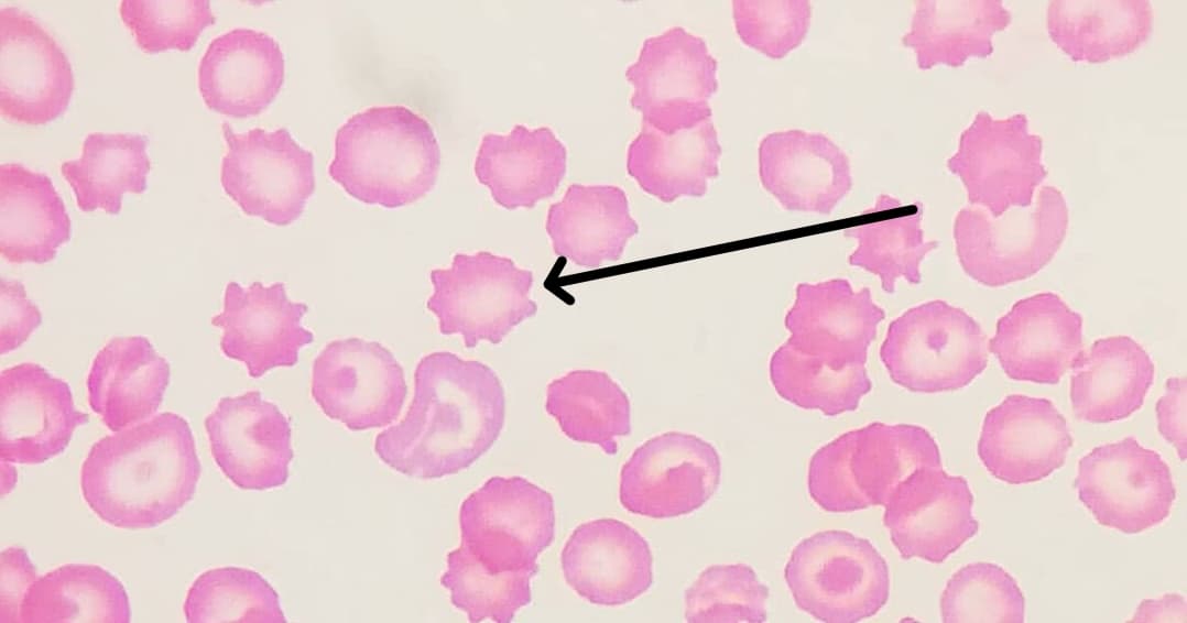
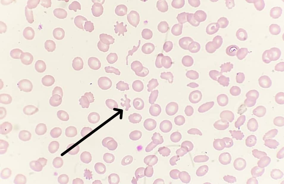
◉ Echinocytes (burr cells)
These are cells with small membrane projections of regular shape and distribution. The number of projections that appear on red blood cells can vary between 10 and 30.
Echinocytes have a central pallor and their projections are more uniform, smaller and blunter than those of acanthocytes.
They can be observed due to increased concentration of fatty acids in plasma (e.g. heparin treatment), in uremia, renal failure, liver disease, hemolyticuremic syndrome and other conditions.
◉ Acanthocytes (Spur Cell)
These are red blood cells that have cytoplasmic projections with a random, heterogeneous and irregular distribution of variable size, some being larger than others.
These cells have no central pallor and their spheroidal shape makes them smaller than normal red blood cells.
Acanthocytes are found in post-splenectomy states, as well as in cases of abetalipoproteinemia and advanced liver disease.
◉ Blister cell
They are caused by the formation of bubbles inside the cell. The bubbles are formed by fusion of the inner cell membrane, causing hemoglobin to concentrate on only one side of the red blood cell.
Red blood cells may have more than one vacuole or bubbles which, when ruptured or phagocytosed, give rise to keratocytes or schistocytes.
◉ Pycnocytes
Pycnocytes are red blood cells that generally have an elongated shape and whose hemoglobin is retracted to the sides of the cell, leaving the central region of the cell empty, without hemoglobin.
◉ Mushroom-shaped red blood cells (pincer cells)
These are red blood cells with cytoplasmic projections, resembling pincer-shaped cells.
◉ Conclusion
In conclusion, the study of the shape of red blood cells is essential for the diagnosis, monitoring and management of patients with hematological disorders.
Continued advances in this field will improve our understanding of the mechanisms underlying red blood cell-related diseases and develop new therapeutic strategies for affected patients.